Fetal Surgery Program
at Hasbro Children's Hospital

Fetal Surgery in Rhode Island
There is a growing branch of pediatric surgery that specializes in treating birth defects while a fetus is still in the womb. This complex surgical subspecialty is sometimes referred to as in-utero surgery or prenatal surgery.
It is used to treat common congenital abnormalities such as twin-to-twin transfusion syndrome, a condition that occurs in 20% of identical twins, and if severe and left untreated, can lead to the death of both twins.
What is In-Utero Spina Bifida Surgery?
Fetal surgery is also indicated in less common conditions, such as spina bifida, in which the baby’s spinal cord doesn’t close properly during prenatal development. Your doctor may recommend fetal surgery if the spina bifida, or myelomeningocele (MMC), is open and the lower spinal is exposed to amniotic fluid while in the uterus.
During an in-utero spina bifida procedure, a team of surgeons makes an incision over the mother’s abdomen and the uterus, partially exposing the fetus. Using microscopic instruments, the defect in the spinal cord is repaired, and the fetus is returned inside the womb.
In some cases of spina bifida and other severe birth defects, it may be preferable to perform fetal surgery rather than wait until the baby is born and then perform an operation. Check with your doctor to determine if prenatal surgery is better for you and your child.
Fetal surgery is also used to remove rare tumors, repair holes in the diaphragm, or remove very large lung lesions.
To schedule an appointment with us or to learn more about this branch of surgery, please call the Fetal Treatment Program of New England at (401) 228-0559, and ask for Debra Watson-Smith, RN, Fetal Treatment Program Director.
BSA Fetal Surgery Specialists
Video: First Successful In-Utero Spina Bifida Surgery in the Northeast
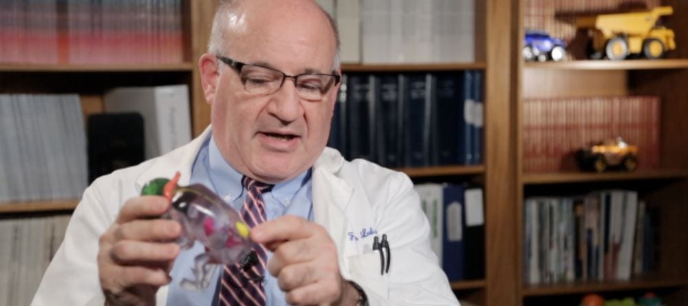
Dr. Francois I. Luks of Brown Surgical Associates’ pediatric surgery division is recognized for his work on the first and only successful in-utero spina bifida surgery performed in New England, right here in Providence, Rhode Island.
In a fetus with this birth defect, a portion of the small intestine called the duodenum becomes restricted by the abnormal growth of pancreatic tissue that forms a ring around the duodenum. This restriction can lead to intestinal blockage, pancreatitis, ulcers, and jaundice.
For more information, visit the American Pediatric Surgical Association website.
This congenital abnormality involves the presence of one or more cysts (fluid-filled sacs) within the bile ducts, the tubes that carry bile from the liver to the small intestine to aid digestion. The cysts may obstruct the ducts, leading to jaundice and an enlarged liver.
For more information, visit the American Pediatric Surgical Association website.
Cloacal exstrophy describes the failure of the lower abdominal wall to develop, leaving critical inner structures such as the bladder and intestines exposed.
For more information, visit the American Pediatric Surgical Association website.
The diaphragm is a muscle that sits below the lungs and aids breathing. It normally contracts with each inhale and relaxes with each exhale, thereby helping air get into and out of the lungs.
In a condition known as diaphragmatic eventration, the diaphragm fails to contract due to a lack of muscle or improper nerve function. This causes the diaphragm to be abnormally positioned (too high up), where it may press against the lungs and make breathing difficult.
For more information, visit the American Pediatric Surgical Association website.
A diaphragmatic hernia is an abnormal opening in the diaphragm – the thin, dome-shaped muscle located just under the lungs, separating the abdomen and chest. It contracts and relaxes rhythmically with every breath you take. This helps to draw air into the lungs during inhalation and expel air from the lungs with exhalation.
When a diaphragmatic hernia is present in a fetus, organs within the abdomen such as the stomach, intestine, liver, and spleen, may move into the chest cavity. It usually occurs with impaired lung development. Other complications may develop as well, such as breathing problems after birth (pulmonary hypoplasia).
Abnormal development of a fetus’s digestive tract can result in:
- Esophageal atresia – an abnormal closure within the esophagus, the swallowing tube connecting the throat and the stomach.
- Tracheo-esophageal fistula – an abnormal connection between the trachea (windpipe) and esophagus, which can cause stomach acid to enter the lungs.
EAs and TEFs tend to occur together.
For more information, visit the American Pediatric Surgical Association website.
With this condition, a hole in the abdominal wall allows the intestines or other abdominal organs to protrude from a fetus’s body. This exposes the organs to amniotic fluid while still in the womb, and potentially to trauma, infection, and dehydration after birth.
With gastroschisis, the hole occurs to the side of where the umbilical cord connects to the fetus. With a similar condition, called omphalocele, the hole occurs at the belly button itself.
A sacrococcygeal teratoma (SCT) is a tumor, usually benign, located at the base of the coccyx (tailbone) in a developing fetus. Depending on the size of the tumor, it could affect proper development of the hips and may obstruct the bowels or urinary tract. This birth defect occurs more often in females than males.
For more information, visit the American Pediatric Surgical Association website.
When prenatal identical twins share a single placenta (monochorionic twins), there could be an imbalance in blood flow known as twin-to-twin transfusion syndrome.
In cases of TTTS, one fetus may receive insufficient amounts of the placenta’s blood supply, which impacts fetal development and can cause in-utero death. The fetus receiving too much of the placenta’s blood supply is at risk of heart failure due to an inability to process the excess fluid.
The urachus is a canal that joins the bladder of a fetus to the umbilical cord. It usually closes down near the end of pregnancy. However, if it doesn’t, a cyst or sinus may develop, which can lead to urine drainage from the belly button.
For more information, visit the American Pediatric Surgical Association website.
In this congenital malformation, blood vessels form a ring around the trachea (windpipe) and esophagus (swallowing tube), which can restrict both passageways.
For more information, visit the American Pediatric Surgical Association website.
Fetal Surgery Resources
- Fetal Treatment Program of New England
- Fetal Medicine Program at Brown
- North American Fetal Therapy Network (NAFTNET)
To schedule an appointment with us please call (401) 228-0559.


